

Clinical case gallery
Explore selected clinical photographs presented for general educational and informational purposes. Clinical cases vary, and images alone cannot establish a diagnosis or determine the appropriate treatment for an individual.
Content Advisory: Clinical Images
This page contains clinical and surgical photographs, including graphic medical images that some viewers may find distressing. Viewer discretion is advised. The images are presented for general educational and informational purposes and should not be used for self-diagnosis or as a substitute for professional medical assessment.
If you prefer not to view graphic medical content, you may leave this page and explore our other services.
Educational Cases
Selected clinical & surgical cases
Each surgical photograph is blurred by default to respect viewer comfort. Click 'View clinical image' to reveal an individual case, or use 'Hide image' to return to the blurred preview.
Graphic Surgical Content
Click below to reveal this clinical photograph
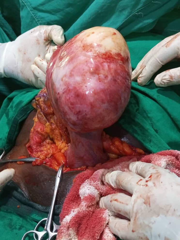
Surgical Case 1
Intraoperative surgical exposure during a complex gynaecological procedure. Presented for educational and clinical documentation purposes.
Graphic Surgical Content
Click below to reveal this clinical photograph
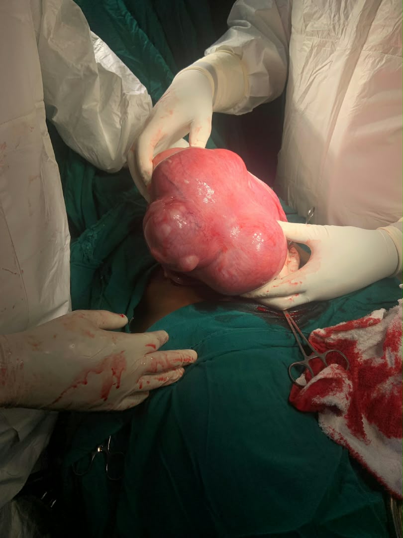
Surgical Case 2
Intraoperative myomectomy / surgical resection demonstrating large uterine fibroid mass during surgery.
Graphic Surgical Content
Click below to reveal this clinical photograph
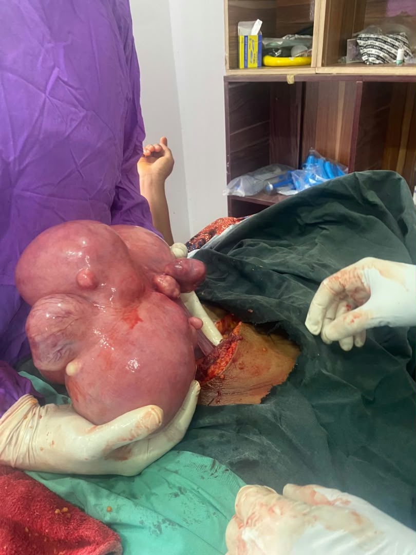
Surgical Case 3
Intraoperative presentation of multi-lobulated uterine mass prior to surgical excision by the gynaecological surgical team.
Graphic Surgical Content
Click below to reveal this clinical photograph
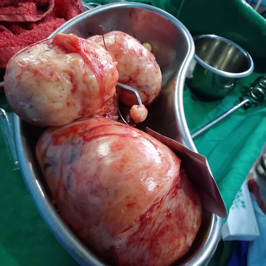
Surgical Case 4
Excised specimen: post-myomectomy resected fibroid masses presented in receiver dish following successful surgical removal.
Graphic Surgical Content
Click below to reveal this clinical photograph
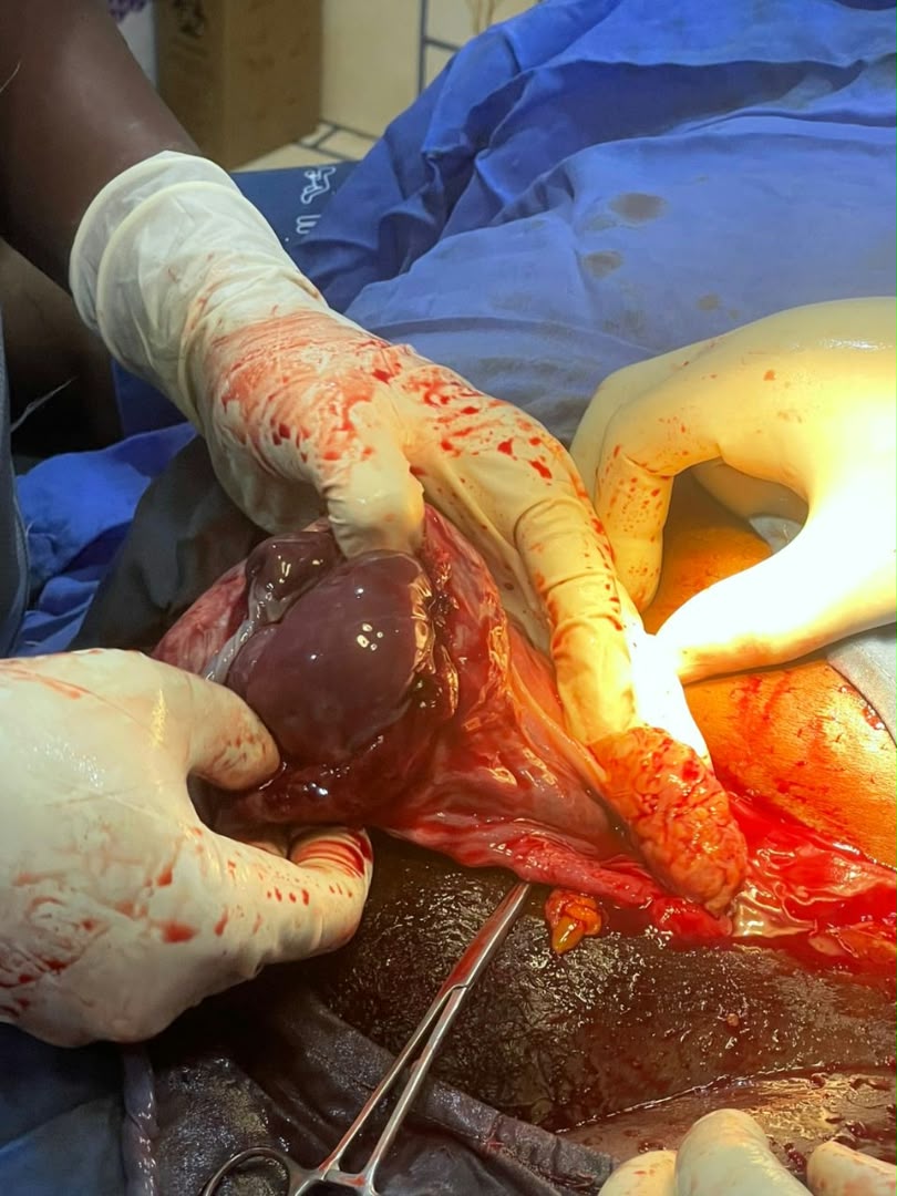
Surgical Case 5
Intraoperative surgical view displaying operative dissection and clinical anatomy during surgical intervention.
Graphic Surgical Content
Click below to reveal this clinical photograph
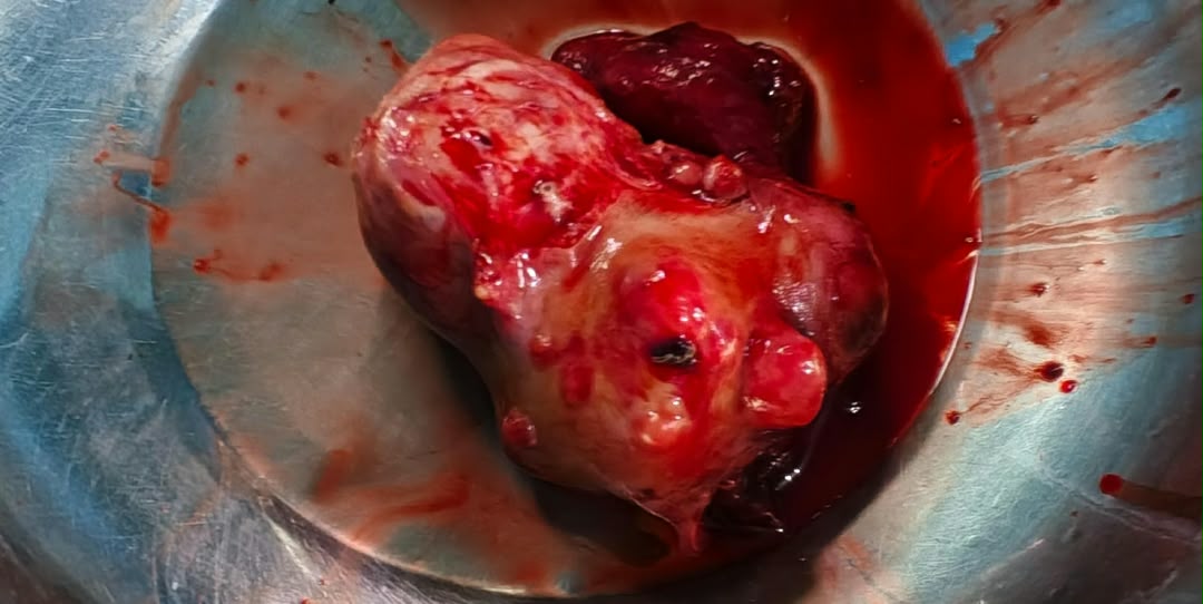
Surgical Case 6
Excised clinical specimen: resected pelvic mass in surgical receiver dish demonstrating macroscopic pathological architecture.
Graphic Surgical Content
Click below to reveal this clinical photograph
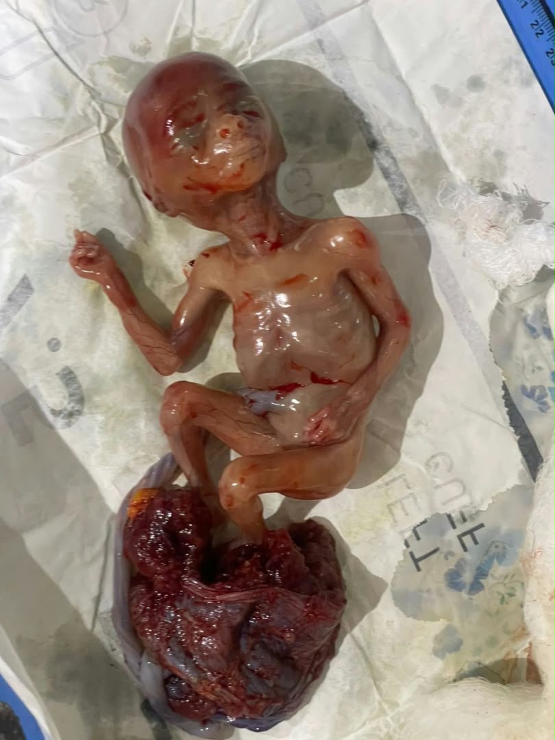
Clinical Case 7
Obstetric clinical presentation: delivered gestational specimen with intact placenta, presented for clinical pathology and educational documentation.
Graphic Surgical Content
Click below to reveal this clinical photograph
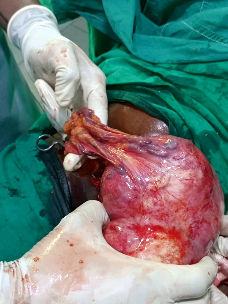
Surgical Case 8
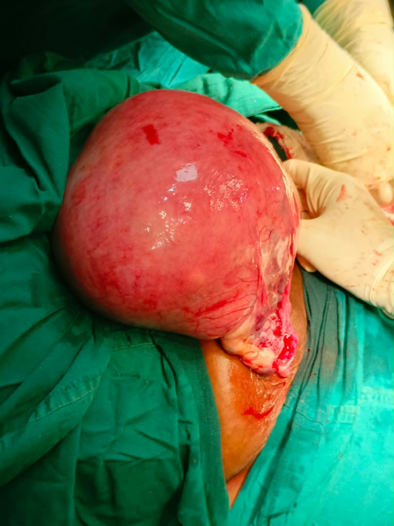
Intraoperative mobilization of large vascularized pelvic mass demonstrating vascular pedicle and surgical dissection field.
Graphic Surgical Content
Click below to reveal this clinical photograph
Surgical Case 9
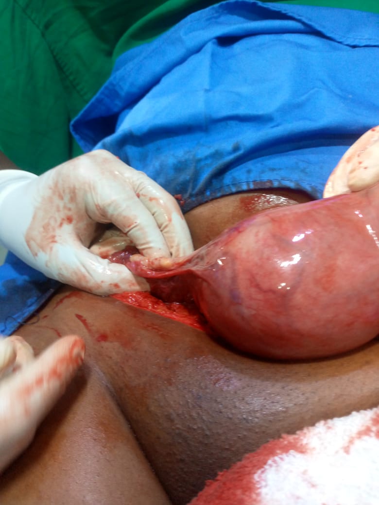
Intraoperative presentation of large smooth-walled uterine mass during exploratory laparotomy and gynaecological resection.
Graphic Surgical Content
Click below to reveal this clinical photograph
Surgical Case 10
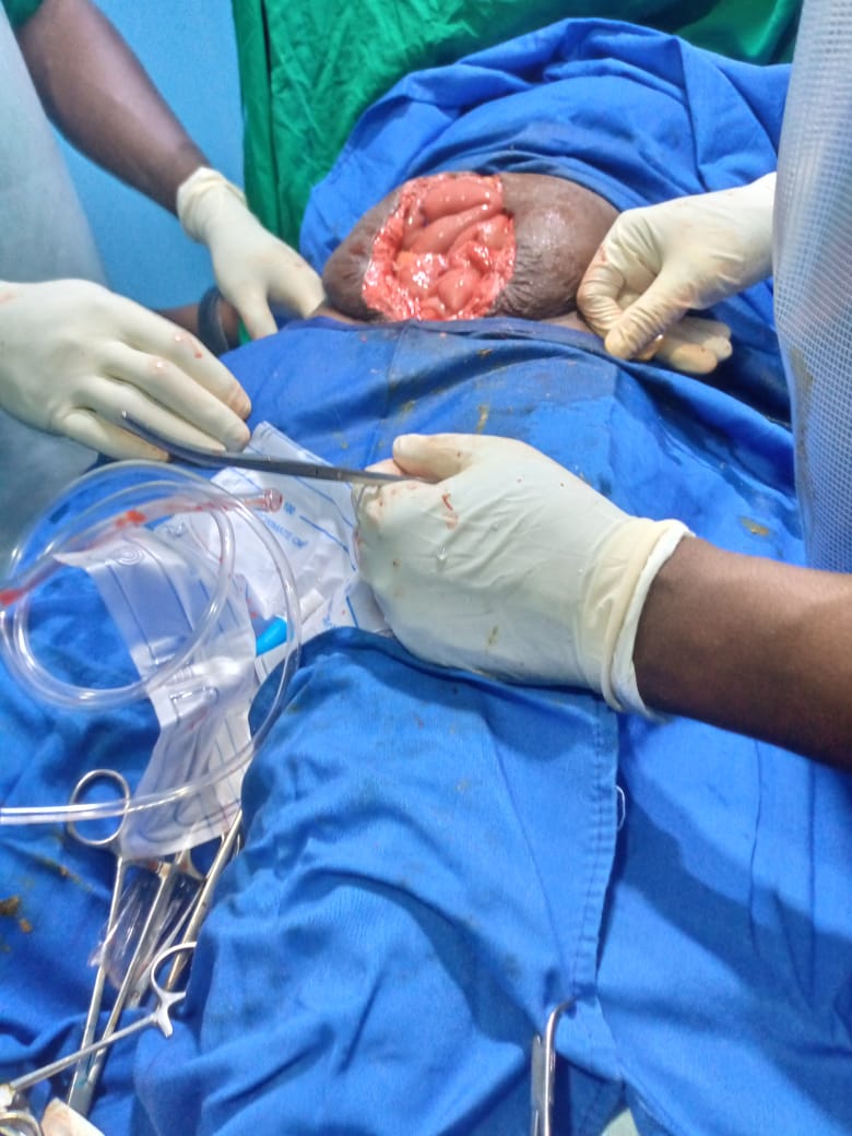
Intraoperative surgical exposure illustrating operative access, tissue margins and haemostasis in gynaecological pelvic surgery.
Graphic Surgical Content
Click below to reveal this clinical photograph
Surgical Case 11
Intraoperative abdominal surgical exposure demonstrating operative field and visceral mobilization under sterile surgical protocol.
Graphic Surgical Content
Click below to reveal this clinical photograph
Surgical Case 12
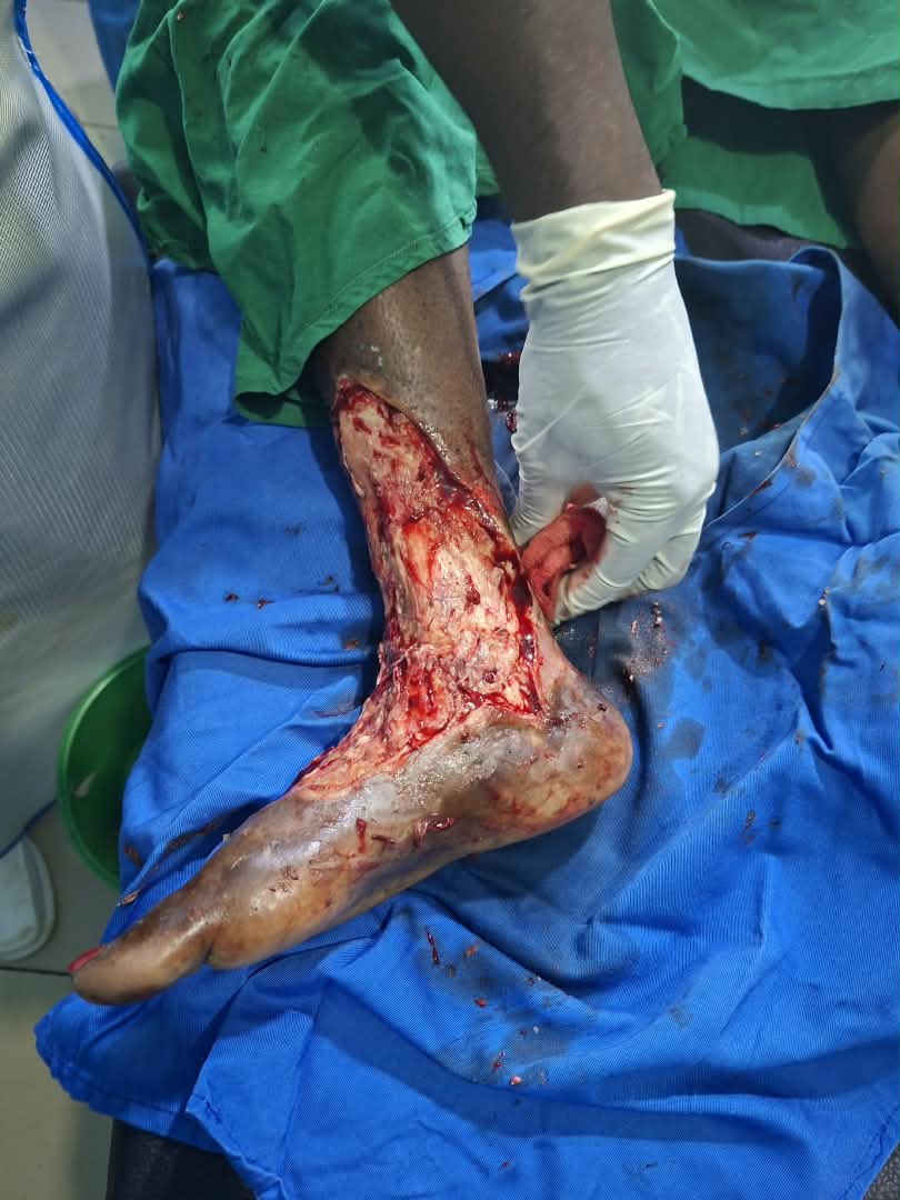
Surgical wound management and soft-tissue debridement of distal lower extremity defect prior to reconstructive intervention.
Graphic Surgical Content
Click below to reveal this clinical photograph
Surgical Case 13
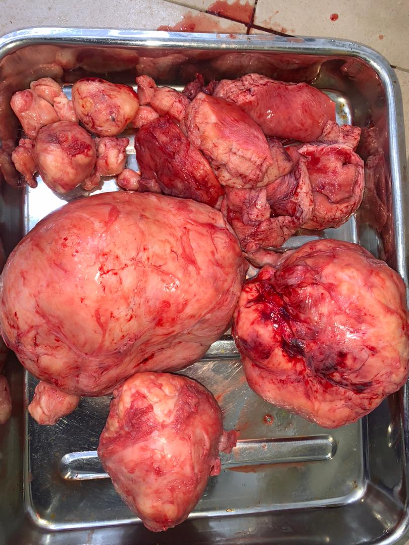
Excised surgical specimen: multiple resected uterine fibroid masses following successful myomectomy, displayed in receiver tray.
Educational disclaimer
The clinical images on this page are provided for general educational and informational purposes only. They do not constitute medical advice and must not be used for self-diagnosis or as a substitute for professional medical assessment. Individual cases vary, and images alone cannot determine the appropriate treatment for any person. For personal medical advice, please contact the centre to arrange a consultation.